Itchy skin and ear problems in dogs: signs to watch and safe first steps
What to look for, why the causes differ, and safe first steps before your dog sees a vet.
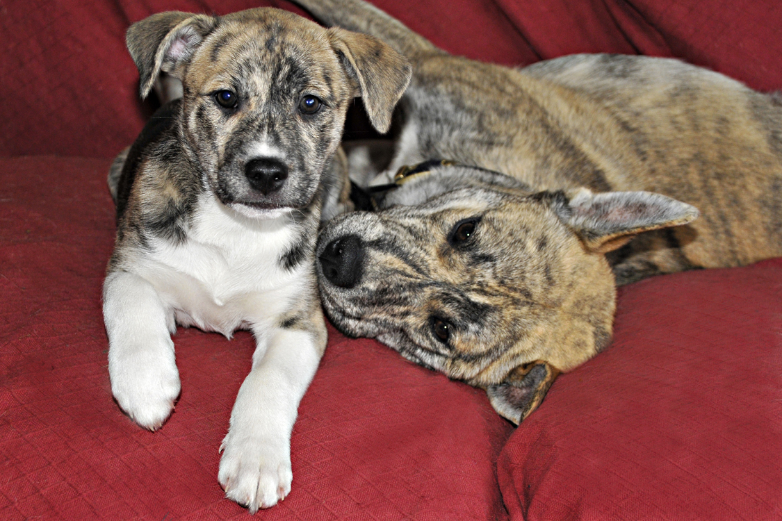
Scratching, licking and head shaking are some of the most familiar things dogs do, so it can be hard to know when they mean something. Skin and ear problems are also among the most common reasons dogs see a vet in the UK. This guide covers what you can usefully notice at home, why the same itch can have very different causes, and which first steps help rather than make things worse.
The short version
- Skin and ear problems are very common in UK dogs. Ear inflammation (otitis externa) was the single most frequently recorded disorder in one large study of English vet practice records. [1, 3]
- Itch is a sign, not a diagnosis. Fleas and other parasites, allergies, food reactions, infections and ear-canal factors can all look similar, and several often happen at once. [4, 7]
- Skin and ear infections in dogs are usually secondary to something else, so the underlying cause matters as much as the infection. [5, 12]
- Do not put cotton buds or anything else into your dog’s ear canal. Do not use leftover drops, someone else’s medicine or a home remedy. [3, 5, 6]
- Write down what you see, take photos and book a vet appointment. A sudden head tilt or loss of balance, a hot swollen ear flap, broken or spreading sore skin, or a dog who seems unwell in themselves needs prompt veterinary attention. [13, 14]
How common are skin and ear problems?
Very common. In a study of the records of 3,884 dogs seen at 89 English veterinary clinics, otitis externa was the most frequently recorded disorder, affecting 10.2% of the dogs. The skin and its related structures (the “integument”) was the body system most often affected. That grouping covers many different skin problems, not just itch. [1]
A later VetCompass study of 22,333 UK dogs under primary veterinary care in 2016 found that 7.3% were diagnosed with otitis externa during the year. [3] An earlier observational survey of UK general practice found that about one in five small-animal consultations involved a skin problem. In dogs, itch (pruritus) was the most common reason, and otitis, skin infection, anal sac impaction, flea infestation and atopic dermatitis were the most common final diagnoses. [2]
These figures come from what vets recorded, so they describe how often problems are diagnosed, not every dog that is mildly itchy at home.
What you might notice
None of these signs tells you the cause on its own, but together they give your vet a much clearer picture.
Skin
- Scratching, licking, chewing or rubbing, including rubbing the face along the carpet or furniture.
- Licking or chewing the paws. The feet are commonly affected in both environmental and food allergies. [7, 10]
- Redness, small bumps or spots, especially on the belly, armpits, groin, face, ear flaps or feet. [7]
- Hair loss, broken hairs or thinning patches where your dog has been licking or scratching. [7]
- Thickened or darkened skin, crusts, scale or a greasy, smelly coat. These tend to appear when itch and inflammation have been going on for a while or an infection has developed. [7, 12]
- Itch with no visible change at all. Early on, allergic itch can happen before anything is visible on the skin. [7]
Ears
- Head shaking or scratching at one or both ears.
- Odour from the ear, or discharge that may be waxy, dark, yellow or bloody.
- Redness or swelling of the ear flap or the visible opening of the canal.
- Pain: flinching, crying or pulling away when the ear or head is touched.
- A swollen, soft or fluid-filled ear flap, usually on the inner surface. This can be an aural haematoma (see below). [13]
- Head tilt, stumbling or circling. These point to the balance system, which needs a vet to assess. [14]
Ear disease can also cause long-term pain and some hearing loss, which owners and vets may not notice as easily as a smell or discharge. [3]
Why the causes differ
Vets often think about skin and ear disease in layers. There is usually a primary cause that starts the problem, secondary infection with bacteria or yeast that takes advantage of inflamed skin, predisposing factors such as ear shape, and, in long-running cases, perpetuating changes such as a narrowed ear canal that keep it going. [5, 6] That is why two dogs with the same-looking ear can need quite different care.
Parasites, including fleas. Fleas, mites (including ear mites and the mites that cause mange), lice and others are on the list of things vets rule out in an itchy dog. [7] Some dogs become allergic to flea bites, so a few fleas can cause a lot of itch. Guidance stresses telling simple flea infestation apart from true flea allergy, and says that clearing fleas from both the dog and its surroundings is essential when allergy is present. [11] You may not see fleas at all: vets check for fleas or flea dirt with a comb, and flea pupae can survive for months. [7]
Environmental allergies (atopic dermatitis). Canine atopic dermatitis is a common, genetically predisposed, itchy, inflammatory skin disease. [7] It often affects the face, inner ear flaps, belly, armpits, groin, around the bottom and the lower legs and feet, although the pattern varies between breeds. [7] Itch may be seasonal, for example with pollens, or year-round, for example with house dust mites. [7] There is no single test that diagnoses it. Vets diagnose it by the history and pattern of signs and by ruling out other itchy conditions first. Allergy tests are used after that, mainly to choose allergens for immunotherapy, not to make the diagnosis. [7] Updated international guidelines describe it as a complex condition that needs individual short- and long-term management, with infections and flare factors addressed as well as the itch. [8]
Food reactions. Cutaneous adverse food reactions (often called food allergy) can cause itchy skin, ear disease and repeated skin infections. In dogs they often affect the ears, feet and belly, and can begin at any age. [10] A systematic review found they are uncommon among all dogs seen by vets (around 1 to 2%) but much more common among itchy dogs, with estimates ranging from 9 to 40% in different studies. [9] The only reliable way to identify one is a strict elimination diet trial planned with your vet. Accurate commercial tests are not currently available, and even small amounts of other food, such as crumbs, treats or licking another pet’s bowl, can spoil a trial. [7] Swapping foods on a hunch usually makes things harder to work out.
Infections that follow something else. Staphylococcal bacteria are thought to live on the skin of all dogs, but most dogs never develop a skin infection from them. Infection develops when the skin barrier has already been altered. [12] International antimicrobial guidelines state that bacterial skin infection (pyoderma) is always secondary to an underlying primary cause. [12] The same applies to recurrent ear infections. [5] Many skin lesions that look infected also occur in non-infectious diseases, so vets confirm infection, often by looking at a sample under the microscope, before choosing treatment. [12]
Ear-canal and ear-shape factors. In the UK VetCompass study, breed and ear carriage were the strongest risk factors for otitis externa. Breeds with pendulous ears had 1.76 times the odds, and breeds with V-shaped drop ears 1.84 times the odds, compared with breeds with upright ears. Basset Hounds, Chinese Shar Pei, Labradoodles, Beagles and Golden Retrievers were among the breeds with higher odds. Chihuahuas, Border Collies, Yorkshire Terriers and Jack Russell Terriers had lower odds. [3] In a study of 100 dogs with otitis externa at one veterinary hospital in Greece, allergic skin disease was the most common primary cause (43 dogs), followed by grass awns, or grass seeds, (12) and ear mites (7). In nearly a third, no primary cause was found. Yeast was found in about two-thirds of the dogs, and most cases affected both ears and kept coming back. [4]
Safe first steps at home
The aim at home is to keep your dog comfortable, avoid making things worse and gather information for your vet.
- Look, do not probe. Look at the ear flap and the visible opening of the canal in good light, and notice any smell. Do not insert cotton buds, tissues, fingers or anything else into the canal. Owner applications such as cleaners and cotton tips are listed among things that can irritate the ear and predispose it to inflammation. [3]
- Do not use leftover or borrowed medicines. That includes old ear drops, another pet’s medicine, human creams and anything prescribed for a previous episode. A vet has to see inside the canal and often look at a sample before choosing a product. Different ear problems need different treatment, and repeated courses that do not address the cause can lead to chronic changes and antimicrobial resistance. [5, 6, 12]
- Skip home remedies and ear-cleaning recipes. Do not put homemade mixtures, household products or oils into a sore ear. UK researchers suggest avoiding overzealous ear cleaning. [3] If your vet recommends a routine cleaner, follow their instructions for your dog.
- Check for fleas, but do not self-diagnose. Part the coat over the lower back and base of the tail and look for fleas or black specks of flea dirt. Finding none does not rule out fleas or flea allergy. [7]
- Check feet and ears after walks in summer and autumn, especially for grass seeds caught between toes or in long ear hair. In the study of 100 dogs, grass awns were the primary cause in 12, particularly in sudden (acute) cases. [4] If you think something is inside the ear canal, do not try to remove it. See your vet.
- Keep a short diary and take photos. Note where your dog itches, when it started, whether it comes and goes with the seasons, what they eat (including treats and chews), any parasite treatment and dates, and any other pets or people with itchy skin. Seasonal versus year-round patterns are useful to your vet. [7]
- Contact your vet promptly if the skin is breaking. Otherwise, gentle distraction, such as a sniffy walk, can help an itchy dog while you wait for an appointment.
Prevention and everyday care
No routine prevents every skin or ear problem, especially when allergies or breed shape are involved. These habits keep the odds in your favour and help you spot changes early.
- Parasite control agreed with your vet, for all pets in the household. With flea allergy, clearing fleas from the surroundings matters as well as from the dog. [11] Your vet can advise on a plan that suits your dog’s lifestyle.
- Regular grooming and handling so you notice redness, smells, lumps or hair loss early, and so your dog is used to having their ears and paws looked at.
- Gentle ear care for dogs with floppy or hairy ears. Look at the ears weekly and wipe only the visible outer part with a dry, soft tissue if it is dirty. The UK otitis researchers give this kind of cautious, low-harm approach as an example of preventive care. [3]
- Keep a stable diet and do not experiment. If your vet suspects a food reaction, the diet trial needs to be strict, so unplanned changes beforehand make it harder to interpret. [7]
- For known allergic dogs, follow the long-term plan. Atopic dermatitis is usually managed over a lifetime, with flare factors and infections addressed as well as the itch. Your vet will tailor this, because no single treatment suits every dog. [8]
When to see your vet
Book a vet appointment if your dog:
- Has been scratching, licking, chewing or head shaking for more than a few days, or keeps coming back to it.
- Has an ear that smells, has discharge, looks red or is sore to touch.
- Has hair loss, spots, crusts, scaling, a greasy coat or darkened or thickened skin.
- Has repeated ear or skin problems, even if they cleared up before.
- Is a puppy or young dog with ongoing itch, or a dog whose itch is getting worse despite treatment.
Seek prompt veterinary attention, the same day if you can, if you notice:
- A sudden head tilt, loss of balance, stumbling, circling or flickering eye movements. These are signs of vestibular (balance-system) disease, which needs a vet to work out where the problem is and what is causing it. [14]
- A swollen, hot or painful ear flap. An aural haematoma usually appears as a swelling on the inner surface of the ear flap, filled with fluid. It can be painful. In UK primary-care records, more than half of dogs with one also had otitis externa recorded. [13]
- Severe ear pain, such as crying out or not letting you touch the head.
- Broken, raw, oozing or bleeding skin, or an area that is spreading quickly.
- Swelling, pain, discharge or ulcers in the skin. These can go with deeper skin infections. [12]
- A dog who seems unwell in themselves, for example quiet or lethargic, off their food, or with skin or ear signs alongside other illness.
If your dog collapses, has difficulty breathing or develops sudden facial swelling, seek emergency veterinary help immediately. These last points are general safety precautions, not findings from a particular study.
What the evidence can, and cannot, tell us
How often. The UK frequency figures come from veterinary records (VetCompass) and an observational survey of practice consultations. They show what is diagnosed and recorded in primary care, not every case. Some mild problems never reach a vet, and records vary in detail. [1, 2, 3]
Risk factors are associations. Breed and ear-shape findings are from observational data. They show which dogs are more likely to be diagnosed, not that ear shape alone causes disease, and breed and ear shape are hard to separate. Many dogs in higher-risk breeds never have a problem. [3, 13]
Causes come from smaller or specialist samples. The breakdown of primary causes of otitis comes from 100 dogs at one university clinic in Greece. That may differ from typical UK first-opinion cases. [4] Food-reaction figures vary widely between studies because the populations studied differ. [9, 10]
Guidelines combine evidence and expert judgement. The ICADA and ISCAID documents are professional guidelines. They review trials where available, but some recommendations rest on expert consensus. [7, 8, 12]
Some of the papers behind this guide were available in full and others only as abstracts; the claims here are limited to what those texts actually say. Some home-care cautions, such as not putting objects into the ear canal or using leftover medicines, follow from the evidence on how ear and skin disease develop, rather than from trials that tested those exact actions.
General information only, not a substitute for veterinary advice. This guide cannot examine your dog, diagnose a skin or ear problem or recommend a treatment. It deliberately does not name medicines or doses.
Sources
- O’Neill DG, Church DB, McGreevy PD, Thomson PC, Brodbelt DC (2014). Prevalence of disorders recorded in dogs attending primary-care veterinary practices in England. PLoS ONE 9(3):e90501. doi:10.1371/journal.pone.0090501. Retrospective records study.
- Hill PB, Lo A, Eden CAN, Huntley S, Morey V, Ramsey S, Richardson C, Smith DJ, Sutton C, Taylor MD, Thorpe E, Tidmarsh R, Williams V (2006). Survey of the prevalence, diagnosis and treatment of dermatological conditions in small animals in general practice. Veterinary Record 158(16):533–539. doi:10.1136/vr.158.16.533. Observational practice survey.
- O’Neill DG, Volk AV, Soares T, Church DB, Brodbelt DC, Pegram C (2021). Frequency and predisposing factors for canine otitis externa in the UK – a primary veterinary care epidemiological view. Canine Medicine and Genetics 8:7. doi:10.1186/s40575-021-00106-1. Retrospective cohort (VetCompass).
- Saridomichelakis MN, Farmaki R, Leontides LS, Koutinas AF (2007). Aetiology of canine otitis externa: a retrospective study of 100 cases. Veterinary Dermatology 18(5):341–347. doi:10.1111/j.1365-3164.2007.00619.x. Retrospective case series.
- Nuttall T (2023). Managing recurrent otitis externa in dogs: what have we learned and what can we do better? Journal of the American Veterinary Medical Association 261(S1):S10–S22. doi:10.2460/javma.23.01.0002. Narrative review.
- Paterson S (2016). Topical ear treatment – options, indications and limitations of current therapy. Journal of Small Animal Practice 57(12):668–678. doi:10.1111/jsap.12583. Narrative review.
- Hensel P, Santoro D, Favrot C, Hill P, Griffin C (2015). Canine atopic dermatitis: detailed guidelines for diagnosis and allergen identification. BMC Veterinary Research 11:196. doi:10.1186/s12917-015-0515-5. ICADA professional guidelines.
- Mueller RS, Banovic F, Bensignor E, Santoro D, Nuttall T (2026). Treatment of canine atopic dermatitis: updated guidelines from the International Committee on Allergic Diseases of Animals (ICADA). Veterinary Dermatology, published online 28 September 2026 (volume and pages not yet assigned). doi:10.1111/vde.70128. ICADA professional guidelines.
- Olivry T, Mueller RS (2017). Critically appraised topic on adverse food reactions of companion animals (3): prevalence of cutaneous adverse food reactions in dogs and cats. BMC Veterinary Research 13:51. doi:10.1186/s12917-017-0973-z. Systematic review.
- Olivry T, Mueller RS (2019). Critically appraised topic on adverse food reactions of companion animals (7): signalment and cutaneous manifestations of dogs and cats with adverse food reactions. BMC Veterinary Research 15:140. doi:10.1186/s12917-019-1880-2. Systematic review.
- Carlotti DN, Jacobs DE (2000). Therapy, control and prevention of flea allergy dermatitis in dogs and cats. Veterinary Dermatology 11(2):83–98. doi:10.1046/j.1365-3164.2000.00204.x. Narrative review.
- Loeffler A, Cain CL, Ferrer L, Nishifuji K, Varjonen K, Papich MG, Guardabassi L, Frosini SM, Barker EN, Weese JS (2025). Antimicrobial use guidelines for canine pyoderma by the International Society for Companion Animal Infectious Diseases (ISCAID). Veterinary Dermatology 36(3):234–282. doi:10.1111/vde.13342. Professional guidelines based on a systematic literature review.
- O’Neill DG, Lee YH, Brodbelt DC, Church DB, Pegram C, Halfacree Z (2021). Reporting the epidemiology of aural haematoma in dogs and proposing a novel aetiopathogenetic pathway. Scientific Reports 11:21670. doi:10.1038/s41598-021-00352-0. Retrospective cohort (VetCompass).
- Rossmeisl JH Jr (2010). Vestibular disease in dogs and cats. Veterinary Clinics of North America: Small Animal Practice 40(1):81–100. doi:10.1016/j.cvsm.2009.09.007. Narrative review.
Evidence checked 10 November 2026. These sources inform the content; their authors have not reviewed or endorsed this article.